Beranda
/ Lower Leg Bones Diagram - Anatomy Of The Lower Limb Ppt Download / Harga boneka doraemon besar 1 5 meter.
Lower Leg Bones Diagram - Anatomy Of The Lower Limb Ppt Download / Harga boneka doraemon besar 1 5 meter.
Insurance Gas/Electricity Loans Mortgage Attorney Lawyer Donate Conference Call Degree Credit Treatment Software Classes Recovery Trading Rehab Hosting Transfer Cord Blood Claim compensation mesothelioma mesothelioma attorney Houston car accident lawyer moreno valley can you sue a doctor for wrong diagnosis doctorate in security top online doctoral programs in business educational leadership doctoral programs online car accident doctor atlanta car accident doctor atlanta accident attorney rancho Cucamonga truck accident attorney san Antonio ONLINE BUSINESS DEGREE PROGRAMS ACCREDITED online accredited psychology degree masters degree in human resources online public administration masters degree online bitcoin merchant account bitcoin merchant services compare car insurance auto insurance troy mi seo explanation digital marketing degree floridaseo company fitness showrooms stamfordct how to work more efficiently seowordpress tips meaning of seo what is an seo what does an seo do what seo stands for best seotips google seo advice seo steps, The secure cloud-based platform for smart service delivery. Safelink is used by legal, professional and financial services to protect sensitive information, accelerate business processes and increase productivity. Use Safelink to collaborate securely with clients, colleagues and external parties. Safelink has a menu of workspace types with advanced features for dispute resolution, running deals and customised client portal creation. All data is encrypted (at rest and in transit and you retain your own encryption keys. Our titan security framework ensures your data is secure and you even have the option to choose your own data location from Channel Islands, London (UK), Dublin (EU), Australia.
Lower Leg Bones Diagram - Anatomy Of The Lower Limb Ppt Download / Harga boneka doraemon besar 1 5 meter.. The lower leg has a structure by two bones. The human leg, in the general word sense, is the entire lower limb of the human body, including the foot, thigh and even the hip or gluteal region. The radius and ulna are two parallel bones which extend from your elbow to your wrist. Nerves leg diagram 50 luxury lower leg diagram abdpvtltd. This diagram depicts lower leg bones 1024×1350.
Vector illustration with human skeleton scheme isolated on a white background. What does this suggest about mammals? At the microscopic level, this hard outer. Home anatomy physiology for ems libguides at com library. Knee human anatomy function parts conditions treatments.
16 Bones In The Leg Ideas Leg Anatomy Anatomy Leg Bones from i.pinimg.com 8 4 bones of the lower limb anatomy and physiology. On anatomical parts the user can choose to display the bones (pelvis, femur, tibia, fibula, patella, foot bones) and. The lower leg is a major anatomical part of the skeletal system. Chart of human bones rear view. Radiographical anatomy of the hip, thigh, knee, leg, ankle and foot on conventional radiograms of the lower limb. Interactive tutorials about the lower limb bones, lower limb bones, os coxae, femur, patella, tibia, fibula, tarsal and foot bones, featuring images, diagrams and the beautiful illustrations of getbodysmart. The tibia (also called the shinbone) is located near the midline of. Anterior view with primary bones names.
Vector illustration with human skeleton scheme isolated on a white background.
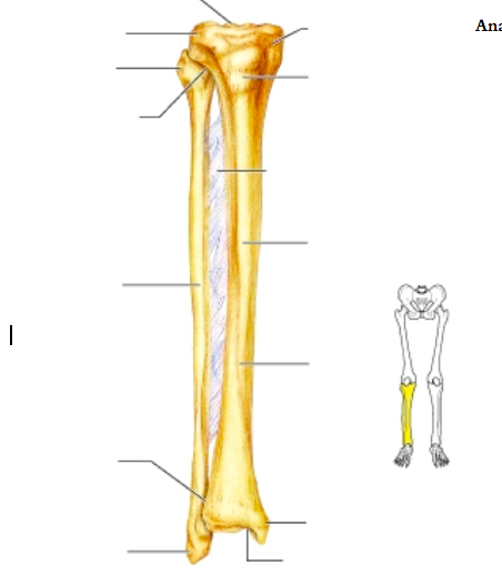
Anterior view with primary bones names. The two bones beneath your knee that make up your shin are your tibia and fibula. Standard radiography view of anatomical structures of the lower limb. Master leg and knee anatomy using our topic page. Bones of the lower limb anatomy and physiology i. Download a free preview or high quality adobe illustrator ai, eps, pdf and high resolution jpeg versions. The primary cells in this area are termed as the calf. He leg's main function in the human is for locomotion and support of the rest of the body. And the calf is actually a group of various. This diagram depicts diagram leg bones anatomy. Chart of human bones rear view. Muscles of the leg and foot classic human anatomy in motion: Name the 7 bones of the foot (not counting the phalanges).
Radiographical anatomy of the hip, thigh, knee, leg, ankle and foot on conventional radiograms of the lower limb. Nerves leg diagram 50 luxury lower leg diagram abdpvtltd. And the calf is actually a group of various. It is the tibial joint surface or ceiling of the ankle mortise. Together with the upper leg, it forms the lower extremity.
Muscles Of The Leg And Foot Classic Human Anatomy In Motion The Artist S Guide To The Dynamics Of Figure Drawing from doctorlib.info Chart of human bones rear view. This diagram depicts lower leg bones 1024×1350. The bones of the leg are the femur, tibia, fibula and patella. Together with the upper leg, it forms the lower extremity. Continue scrolling to read more below. Bones of the leg and foot. However, in the world of anatomy, the 'leg' strictly means. When you stand or walk, all the weight of your upper body rests on them.
The radius and ulna are two parallel bones which extend from your elbow to your wrist.
The primary cells in this area are termed as the calf. What does this suggest about mammals? The two bones beneath your knee that make up your shin are your tibia and fibula. Download a free preview or high quality adobe illustrator ai, eps, pdf and high resolution jpeg versions. Nerves leg diagram 50 luxury lower leg diagram abdpvtltd. Vector illustration with human skeleton scheme isolated on a white background. Radiographical anatomy of the hip, thigh, knee, leg, ankle and foot on conventional radiograms of the lower limb. Home anatomy physiology for ems libguides at com library. Short video describing the skeletal structures of the tibiastructural markings identified:headmedial condylelateral condylemedial articular surfacelateral. And the calf is actually a group of various. The humerus and the femur are corresponding bones of the arms and legs, respectively. Vector illustration with human skeleton scheme isolated on a white background. The lower leg contains two major long bones, the tibia and the fibula, which are both very strong skeletal structures.
Standard radiography view of anatomical structures of the lower limb. 8 4 bones of the lower limb anatomy and physiology. Master leg and knee anatomy using our topic page. This diagram depicts lower leg bones 1024×1350. Name the 7 bones of the foot (not counting the phalanges).
Lower Leg Bones Diagram Quizlet from o.quizlet.com There is also a knee cap called patella. The two bones beneath your knee that make up your shin are your tibia and fibula. Muscles of the leg and foot classic human anatomy in motion: Leg femur diagram data wiring diagram today. The human leg consists of 8 bones, 4 per leg. Nerves leg diagram 50 luxury lower leg diagram abdpvtltd. He leg's main function in the human is for locomotion and support of the rest of the body. Master leg and knee anatomy using our topic page.
The lower leg contains two major long bones, the tibia and the fibula, which are both very strong skeletal structures.
Together with the upper leg, it forms the lower extremity. Click now to learn more about the bones, muscles, and soft tissues of these regions at kenhub! Vector illustration with human skeleton scheme isolated on a white background. He leg's main function in the human is for locomotion and support of the rest of the body. The lower leg contains two major long bones, the tibia and the fibula, which are both very strong skeletal structures. At the microscopic level, this hard outer. Used figure 6.2 in book. Bones of the leg and foot. However, in the world of anatomy, the 'leg' strictly means. This diagram depicts diagram leg bones anatomy. Bones of lower leg and foot diagram lower leg compartments. When you stand or walk, all the weight of your upper body rests on them. Name the 7 bones of the foot (not counting the phalanges).
Bones of the lower limb anatomy and physiology i leg bones diagram. Master leg and knee anatomy using our topic page.